Painting with molecules
Dortmund-based biochemists are able to draw patterns in cells. All that is needed to paint with “molecular ink” is a laser and light sensitive molecules.

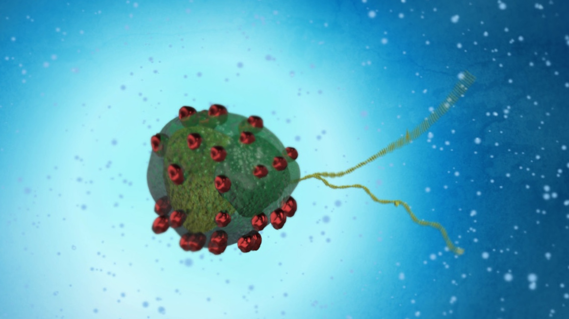
Molecular structures are the building blocks of every organism. But because of their minute size, they are extremely difficult to visualize. A team of Dortmund-based researchers headed by Leif Dehmelt at the Technical University and Yaowen Yu at the Chemical Genomics Center of the Max Planck Society developed a new method to depict the inner workings of a cell. They published their result and their new method entitled “Molecular –Activity Painting” in the renowned journal “Angewandte Chemie“.
Tracing molecular activity
Proteins are criss-crossing the cells for several molecular processes. If theses protein movements could be visualized, it would make it much easier to trace and decipher many of these complex processes. Most difficult to trace and understand have so far been processes occurring close to the cell membrane.
Dortmund-based scientists now managed to devise a new method that is disregards this difficulty. Their new approach of “Molecular- Activity Painting” (MAP) is a new technique that enables the stable trace of signal molecules and their activity close to the cell membrane on a micrometer scale.
Light sensitive molecules are activated by laser-light
In a first step the team of researcher needed to anchor the freely moving parts of the cell membrane. In a second step, specialized light sensitive molecule-systems were introduced into the cells via these anchors. Distinct proteins then bind to these molecule-systems. Subsequently these compounds are activated from the outside by a selective and targeted laser beam – now the “painted” picture is visible.
Depending on the proteins that bind to the molecule-system, distinct molecular processes can be observed. Some proteins even cause muscle-like contractions within the cell, which can now also be visualized. These contracting structures are especially important when it comes to embryogenesis or metastasizing cancerous cells.
The goal of this research project is to track and thereby understand these complex developmental and disease-related processes.
jmr
You might also like

Achieving vanilla flavor through bioeconomy
Microalgae as biohybrid microswimmers