Watching the inner workings of plants
For the first time the distribution and utilisation of the ubiquitous energy currency ATP was visualised in living plants. This may aid future plant breeding schemes.

Adenosine triphosphate (ATP) is the ubiquitous energy currency of all living organisms. Without it there would be no metabolic processes or growth possible – for animal cells as well as plant cells. Headed by the University Bonn an international team of researchers was able to visualise the ATP distribution and utilisation during stressful phases in living plant seedlings. The results were published in the journal „eLIFE“ and could aid future breeding schemes for stress-resistant plants.
Visualising the energy transport
ATP is a chemical molecule that provides universal and immediate energy release. “Our work makes this energy carrier visible,” says Markus Schwarzländer, head of the Emmy-Noether Group at the Institute of Crop Science and Resource Conservation (INRES), University of Bonn. “In living plants, in fact – from the smallest cell organelle up to the complete seedling,” adds his colleague Stephan Wagner. Led by the two INRES biochemists, scientists from Germany, Italy, China, England and Denmark have developed an innovative way of visualizing ATP in the living plant aided by a fluorescent protein. The team used a method developed by Takeharu Nagai from Osaka in Japan, in which ATP binds to a fluorescent protein from a jellyfish. The Japanese researchers had originally used this technique in mammals, and the researchers at the University of Bonn have now optimized it for use in plants. “This method makes it possible to track where and how much ATP is present in the living plants in real time,” says lead author Valentina De Col.
Watching ATP in a living organism
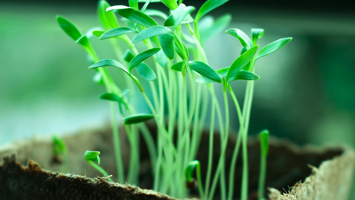
Importantly, previous studies on ATP were only able to provide snapshots. Whole plants or plant parts were ground up before ATP could be quantified. “In contrast, our methodology allows watching the running machine at work”, says Schwarzländer. The “machines” are seedlings of the model plant Arabidopsis thaliana. The researchers investigated tiny cell organelles, such as the cellular powerhouses (mitochondria), as well as entire organs, such as roots and even whole seedlings, under the microscope and with a fluorescence analyzer. Live-imaging showed the distribution of cellular fuel in real time. “With a normal supply of water, air and light, there is less ATP in the roots than, for example, in the green leaves,” reports Wagner.
Insights into how pathogenes intervene
In order to analyse how ATP is distributed and used under stressful conditions, the researchers placed the glowing Arabidopsis seedlings under water to cut them off from their vital oxygen supply. This, however, did not immediately inhibit ATP production. Thus, there have to be different acclimation processes that are activated by the plant in an attempt to protect itself against progressing oxygen shortage and to maintain its energy balance. “A crucial question is now whether these protective programmes can be stimulated to breed plant varieties that cope better with stress”, says Schwarzländer when asked about future opportunities built on these results. Using this new visualisation tool may allow novel insights into how pathogens intervene in the plant energy metabolism and how the symbiosis between roots and specific fungi, called mycorrhiza, works in detail.
jmr


